METALLIC PARTICLES AFTER PHACOMULSIFICATION
Posted by J.R. Villada on October 9, 2015 in Blog | Comments Off on METALLIC PARTICLES AFTER PHACOMULSIFICATION

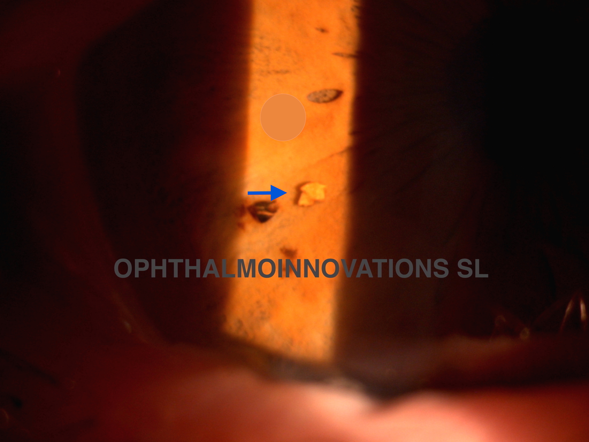
This picture was taken by Julian Tendero, Optometrist from Baviera Clinica in Albacete (Spain). It shows a metallic? foreign body after phacoemulsification. We have reviewed the subject and we here show a resume.
Metallic pieces in the anterior chamber, usually attached to the iris, after phacoemulsification, are very common. The first publication I could find about it comes from Dunbar et al. in 1995 (Dunbar CM, Goble RR, Gregory DW, Church WC. Intraocular deposition of metallic fragments during phacoemulsification: possible causes and effects. Eye (1995) 9:434-436). They studied in the slit-lamp 56 eyes which had undergone routine phacoemulsification and found metallic particles in 86% of the eyes. They studied and photographed twelve phacoemulsification needles and concluded that the cause of the FBs is not the touching of metallic instruments with the vibrating phaco tip, but the wear of the middle part of the needle. They also showed their concern because these particles are not inert and they could attract macrophages that release lysosomal enzymes, collagenase, interleukines and prostaglandines. Does it sound like potential TASS?.
Soon after, Braunstein et al. in 1996 (Braunstein RE, Cotliar AM, Wirostko BM, Gorman BD: Intraocular metallic-appearing foreing bodies after phacoemulsification. J Cataract Refract Surg 1996;22:1247-1250). suggested that touching between the phaco tip and the metallic instruments produces these small pieces that usually stay stuck to the iris. They show their concerns about what will happen to these foreign bodies (FB) in the long term. Also what will happen in case of needing a MRI scan. They mention a paper by Kelsey et al. (Kelsey CA, King JN, Keck GM et al. Ocular hazard of metallic fragments during MR imaging at 0,06T. Radiology 1991; 180:282-283) where the authors state that a metallic FB will not move if it is not bigger than 560 microns).
Davis et (Davis PL, Mastel D. Anterior chamber fragments after phacoemulsification surgery. J Cataract Refract Surg 1998; 24:810-813) had noticed the metallic particles back in 1988 and also had heard the explanations of a spokesperson from a manufacturing company, who recommended that all new needles were run for two minutes before use. They think that the particles are attached to the new phaco needles and become loose during the procedure. They also give information about a report from the SMRI Safety Committee, that suggest that metal fragments in animal eyes must be 3.0 x 1.0 x 1.0 mm before they rotate in the eye.
The Query No: 1857 of the All India Ophthalmological Society, published in 2013, asks about the origin of these metallic particles after phacoemulsification. Each of the six answers gives a different provenance: from the second instrument, from the viscoelastic cannula, from the internal lumen of the I/A tip, phaco tip as it gets older and from the wrench used to tighten the phaco tip. But look at the interesting answer fro Dr Sunil Moreker:
“You touch upon a very important medicolegal topic of foreign body left in eye after surgery which can potentially cause litigation Many general surgeons have faced consumer court actions due to Gossypiboma but eye surgeons have not yet faced the courts in this matter or Metallopiboma Many of these particles which are “metallic appearing” may not be metallic in fact as has been elucidated by Braunstein et al from New york who published the findings in the Journal of Cataract and refractive surgeons Nov 1996 and proved that these metal fragments can be experimentally produced by applying phaco handpiece to a cyclodialysis spatula and hence probably form due to touch between instruments and I believe that reverse chop prevents this touch between instruments Dumbar et al in the journal Eye in 1995 discussed the possible cause and effects of metal in eye and found the pattern of wear and tear at the area under the sleeve and suggested a cavitational erosion Davis et al have in the Journal of Cataract and Refractive Surgeons, Jun 1998, discovered that on new titanium phaco needles scanning electron microscopy found adherent metal on lathed surface but these were not found on used needles suggesting new needles need to be used carefully and also not to use needles more than the recommended usagesKose et al in their paper in Ophthalmologica in 2003 May June , published their finding after they applied USG linear power 100 percent for 5 min continuously, with a new tip and used tip ,and filtered it and studied it on SEM and found metal on the filter used for new tip thus confirming Davis ‘ findings Martines -Toldos et al from spain published in JCRS Sept 1998 that there are irregularities on sinskey hook and phaco tip Arbisser et al identified silver originating from the brazing of irrigation tube as it entered the handpiece and published this in JCRS 2005 Manjunaths et al have discovered a large foreign body in angle of anterior chamber after phacoemulsification and reported it in the journal eye in 2007 Chaudhari M, Agarwala NS and Nayak BK in their paper in JCRS 2013 found metal in 11 eyes during the period June 2010 to Mar 2012 and suggested these to be arising from the wrench used to tighten phaco needle and suggested a plastic wrench or using it carefully but a Storz notice in Health Devices 1993 probably says that undertightening can cause release of iron and can cause siderosis Bausch and lomb had given a public advisory in Jan 2010 that if the wrench is not used properly and if the surgeon is negligent in not washing the tip after tightening even a plastic wrench may cause plastic foreign body in eye and should be looked for and removed immediately ( Reference :- http://www.swissmedic.ch/recalllists_dl/02941/Vk_20100113_05-e1.pdf ) Whatever the cause we stand to face charges of negligence especially if we end up leaving metal in eye after a bausch and lomb advisory telling us that one should inspect the tip and wash it to remove particulate matter and examine the eye at end of surgery to look for these foreign bodies and remove them at that time itself. I use a hand held slit lamp at times when I suspect this It would constitute negligence if one doesn’t suspect and look for them in cases of recalcitrant inflammation post phaco. Removing the metal may be tricky but Christpher king et al In cataract and Refractive surgery today Feb 2006 explain how a dispersive viscoelastic with only soft silicon aspiration line without irrigation line can be used to remove a metal on a multifocal IOL where it will definitely cause glare and other visual complaints ( can forward the pdf) Stethen Shriver et al have shown in Journal of Ultrasound Med in 2005 , how even hand held sonography is highly sensitive and specific in detecting very small metal foreign bodies like what we have detect at Eyeris medical center at Vile Parle, Mumbai CT is known to miss even large fragments of broken chopper tip as reported by Wu et al in British Journal of Ophthalmology 1998 Mathys et al have discovered and reported in JCRS 2008 that the foreign bodies left behind may be a cause of Toxic anterior segment syndrome Stangos et al in American Journal of Ophthalmology 2005 have identified occult anterior chamber foreign bodies post phaco masquerading as chronic recalcitrant post operative inflammation Occult hidden metal in anterior chamber is like a “microbullet” in the patients eye when he or she undergoes an MRI and literature has more than one such case report which describes the dangers In our instruction course at ESCRS London 2006 we demonstrated how metal causes endotheliam loss and how an AC maintainer during phaco or a high flow system will cause more turbulence and cause endothelial loss in presence of these metals Dr Ranjit Maniar in his joint presentation with me in annual conference of Implant Society of India 2007 showed how this damage can be reduced Most eye surgeons end up defending colleagues or undermining the side effect of retained metal in eye but the evidence is catching up and ultimately so will the courts and finally if we leave metal from a wrench in the eye when there is an exisiting communication from a company we are bound to be charged and punished for negligence. So we ought to be very careful, observant, watch our phaco needle tips, sleeves, wrenches and the procedure being followed to tighten the needles , should wash the phaco tip after assembly as adviced by company, and recheck the eye after the surgery so that we can be absolved of any charges of negligence This communication from me is pretty long and I will forward the court cases in this regard whenever anyone asks for it. There is a PIL in honorable courts in India on Titanium release in orthopaedic implants and investigations are on and these metals at times have titanium as a constituent. In particulate form it attracts macrophages as pointed out by Scales et al. There are reports of titanium hypersensitivity and possible role in oncogenesis has been postulated by authors like Black et al and Gillespie et al and Abdallah et al
Thanks for bringing this up for the benefit of all members of AIOS”
So, what do you think?, where are this particles coming from?, are they dangerous?, capable to produce TASS?, a legal problem?.
Dr José R. Villada